

“Iowa friends – listen up. You have a great opportunity because someone in your area is now offering thermography! “MEND” will help you get on the mend! Had I had this technology sooner, perhaps we could have caught the cancer sooner and that’s why I’m such an advocate. “You’re too young to have cancer…. you look well…..” Well guess what? Cancer doesn’t care. It didn’t show in my blood work, and it didn’t show in a sonogram. It was TWO Thermograms that showed things were going on – that finally led to an MRI over a year later… By the time they figured out what was going on – it was stage IV. Never trust “just” one test. If you, or someone you know needs a mammogram – or struggles with disease in their family (Stroke, chronic pain, lyme, auto immune etc) – this is as safe as you get for testing to see where your body is struggling!” –Sara V.
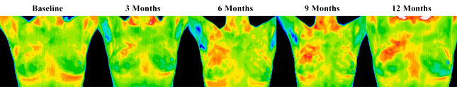
This patient is a 37 year old female whose first baseline thermogram showed a slight hyperthermic asymmetry in the upper right breast. The follow up study showed the pattern had become more well defined & although clinical correlation did not find anything remarkable, it was decided to repeat the exam again in a further 3 months when again significant changes were seen. Mammography was performed at this stage with the thermographic guidance of a locally suspicious area at one o’clock to the right nipple. The mammographic findings were inconclusive and the patient was referred for a repeat mammogram in twelve months. Thermographic monitoring ws continued and at the fifth comparative study at twelve months significant changes were still evident and the hyperthermic asymmetry (temperature differentials) had increased. Immediate further investigation was strongly recommended despite a scheduled mammogram in six months. At the patient’s insistence a repeat mammogram was performed that clearly showed a small 1mm calcification at one o’clock in the right breast. An immediate lumpectomy was performed with good margins and the pathology confirmed as a malignant carcinoma (DCIS). The patient has had stable thermograms for the last two years and is expected to remain healthy.
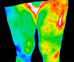
Elderly female patient that had undergone a left hip replacement three months previously. Her continued leg pain raised a suspicion for deep vein thrombosis (DVT). The thermographic findings were not consistent with DVT, but showed a focal area of inflammation that guided a sonographer to a deep abscess near the bone. The abscess was lanced and treated successfully with antibiotics.
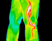
A football player with a stress fracture that was not detected with x-ray. A scintigraph (bone scan) confirmed thermographic findings.
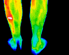
“I actually began to wonder if my pain was real. With all the tests I’d had, surely one should have shown some reason for my pain. With the DITI scan, my doctor, my family, and I can see the cause of my pain. And now I know I’m not going crazy.” This phlebitis was not detected by other tests. Vascular pain and inflammation can be graphically shown with our DITI camera.
The client presented without any symptoms or visible signs of an abnormality. Her thermogram revealed a inflammatory process occurring in the right breast. Referral to a breast specialist and a subsequent biopsy diagnosed inflammatory breast cancer at a very early stage.
